
MRI (Magnetic Resonance Imaging), one of the most advanced and advanced radiological methods used in medical diagnosis, creates images using the natural parameters of the human body. MR devices consist of devices with a magnetic field strength between 0.2 and 3 Tesla MR.
NPISTANBUL Hospital offers high quality image and patient comfort with its large magnet (70 cm) 3 Tesla MRI device, which is available in few centers in our country. Thus, detailed images are obtained.
3 Tesla Technology is designed to obtain images with high resolution and sharpness. The magnet forms the basis of the MR system, and systems with 3 Tesla magnetic field strength increase the diagnostic capabilities of doctors while allowing patients to experience a faster and more detailed examination experience.
Advantages of 3 Tesla MRI
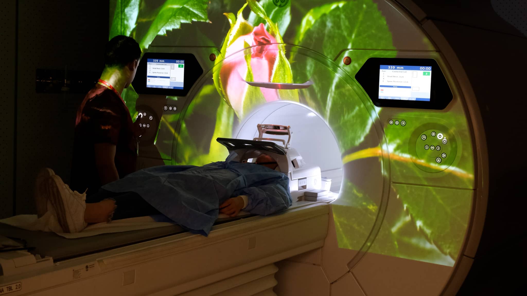
- The spaciousness of the device increases the patient's comfort and adaptability to MRI. It also makes it possible for patients who are afraid of being indoors and overweight to undergo MRI.
- One of the most important factors affecting patient comfort in 3T devices is the high noise level during the examination. Some patients may be very uncomfortable with this. The 3T device has a sound reduction technique that can reduce the sound heard by the patient by up to 40 percent.
- There is also a music system and video broadcast for patients to have a pleasant time during the examination.
- Image quality is high in all shots thanks to a program that prevents image distortion caused by movement, MRI-compatible metal prostheses and implants .
- It represents a special approach to make pediatric patients feel more comfortable and relaxed during their Magnetic Resonance (MRI) scan. This experience aims to make the scanning process easier for children with indoor phobia or anxiety. This special design aims to calm and relax children by distracting them with factors such as exterior and interior ambient effects, special images and music. For example, colored lights or pleasant scenery can be used to distract the child from the screening process. In addition, scanning accompanied by soft music can also help children to calm down.
- Among the methods of application for individuals with special needs, shots are safely performed under sedation anesthesia.
What is the Application Process of 3 Tesla MRI?
- Before the procedure, the patient should not have any metal such as necklaces, earrings, watches and rings. The patient is first asked by the team to remove all metals.
- The patient lies down on the MR device and the lying position according to the area to be imaged is shown to the patient by the team.
- During the procedure, the patient hears and realizes the necessary warnings through the headphones attached to his/her ears. The earphones also allow the patient to be less affected by the sound of the device.
- There is a button in the patient's hand to quickly call the team in case of any problems.
- The acquisition time varies depending on the area to be imaged and the type of disease.
What are the Application Areas of 3 Tesla MRI?
3 Tesla MRI can be applied for imaging organs and systems in different parts of the body. In addition to neurological disorders such as brain tumors, epilepsy, spinal cord diseases, migraine, ear disorders, jaw problems, spine diseases, prostate diseases, breast diseases, joint and connective tissue imaging such as shoulder and knee, 3 Tesla MRI is used in the detection of many problems related to bones, imaging of coronary vessels feeding the heart and whole body imaging.
When and why is it needed?
In cases where other radiological modalities are insufficient in diagnosis or in the presence of findings that cannot be explained by the patient's clinical condition, MRI, one of the advanced radiological examinations that play a leading role in the diagnosis of diseases, is requested.
MRI is among the first tests requested for the evaluation of many organs, such as the brain, which cannot be shown on direct radiographs. Computed Tomography gives more successful images in evaluating bone structures, while MRI gives more successful images in showing soft tissues.
- Advanced Image Techniques
- Advanced MR imaging
- Virtual colonoscopy
- MRI-enterography
- Multiparametric prostate MRI
- MR-fistulography
- Functional MR
- MR spectroscopy (Multivoxel, single voxel and 3D voxel)
- Diffusion tensor imaging (DTI)
- MR perfusion
- MR cisternography
- MR arthroscopy,
- Cartilage T2 mapping imaging studies
NP İSTANBUL Hospital Radiology Clinic also offers color mapping and examination from different angles thanks to the latest technological computer programs.