Standard MRI is an effective and harmless imaging method. One of the most important advantages of MRI is that there is no pain after MRI and there is no need to take any medication that may cause allergies during imaging.
Magnetic Resonance Imaging is a medical technique used to clearly distinguish certain anatomical structures from other structures, to detect and identify differences between healthy and diseased tissues by using radio waves in a strong magnetic field created by large magnets. With this feature, it is a method that can be used safely for diagnostic purposes even in very young babies and pregnant women (it is not preferred in the first trimester of pregnancy unless there is an absolute necessity).
In addition, with the help of MRI compatible ventilator devices in patients who are afraid of being in a closed place, and with MR compatible LED TVs that will keep them immobilized inside in very young children and infants, the desired video etc. movies can be safely shot during the shooting (optionally).
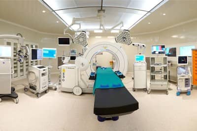
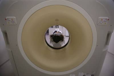
The standard MR system in our hospital is a device with a full neuropack of 1.5 tesla power and includes technological developments. With its electrophysiological use, NPISTANBUL Hospital is the first psychiatric hospital in Turkey to have an MRI. With this device equipped with sufficient and special programs for each region;
- Head region examinations such as brain, eye, inner ear and ear structures, pituitary, jaw joint, brain artery and vein systems,
- Neck structure, larynx, pharynx, pharynx, salivary glands, tongue and surrounding structures,
- Lungs, heart and large vessels associated with the heart,
- Intra-abdominal organs, lower abdomen,
- Spinal pathologies in the neck, back and lumbar region,
- Examination of limbs and joints such as shoulder, arm, elbow, wrist, hand, hip, thigh, knee, leg, ankle and foot
- MR spectroscopy,
- Cranial and abdominal diffusion imaging,
- Perfusion MRI
- MRCP, MR pyelography and MR myelography.
- CSF flow study,
- Kinematic investigations,
- Dynamic tissue (liver, tumor) MR,
- Regional MR angiographic examinations,
Why is an MRI performed?
MRI can be applied for different purposes for different parts of the body. MRI evaluation can be performed in migraine, headaches, neurological disorders, patients with suspected brain tumors, patients with epileptic seizures, patients with eye, ear, jaw joint problems, spine problems, slipped discs and disc herniations, evaluation of joints and ligaments such as shoulders and knees, sports injuries, heart diseases, chest and abdominal internal organ disorders, bone structure disorders.
How is MRI performed?
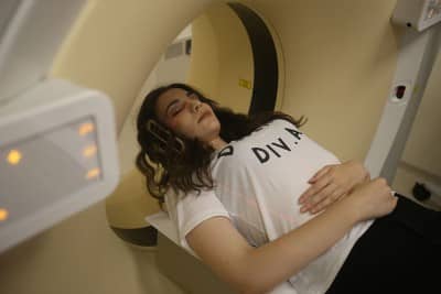
No extra preparation is required for MRI. Unless otherwise warned, the patient can eat their meals and take their medication. The patient is required to fill out a form about his/her medical history for MRI. In addition, the patient must remove any items that may be affected by the magnetic field such as watches, credit cards, metal objects etc. before entering the MRI room. If the bladder is full, there is no harm in urinating before the scan unless otherwise instructed. The examination period usually lasts between 15-45 minutes. During this time, the patient will be asked to remain still. The patient should be aware that the slightest movement will cause distortion in the images. In some cases, specially designed MR contrast agents may be injected to improve image quality and increase the safety of the diagnosis. These drugs will help to clarify the details of the MRI images.
MRI of the brain
It is an important method for detecting chronic nervous system diseases such as brain tumors, strokes, dementia and multiple sclerosis. It is also a good method for evaluating diseases of the pituitary gland, brain vessels, eye and inner ear organs.
It can be difficult, risky and time-consuming to perform biopsies with known methods to obtain information about lesions in the brain. With advanced MR imaging methods, it can be determined whether the suspected lesion in the brain is a tumor or not. Advanced MR imaging methods such as Diffusion MR, DTI MR, Functional MR, Perfusion MR and MR spectroscopy can be used to evaluate the extent, type, metabolic and biochemical structure of the tumor and its relationship with the areas and pathways that enable speech, vision and movement. The data obtained with advanced MR imaging methods enable the determination of treatment approaches.
Safe Roadmap for the Surgeon
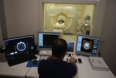
While advanced radiological imaging methods play an important role in diagnosis, they also determine the safe road map for the surgical team that will perform the surgery. The tumor is examined in every aspect using advanced MRI methods before surgery. In particular, the relationship of the tumor with the limb movements, vision and speech centers in the brain is determined in detail. The 3D images obtained are superimposed and uploaded to the "Neuronavigation" device used in the surgery. In this way, the surgical team is given a short and safe road map to reach the tumor.


