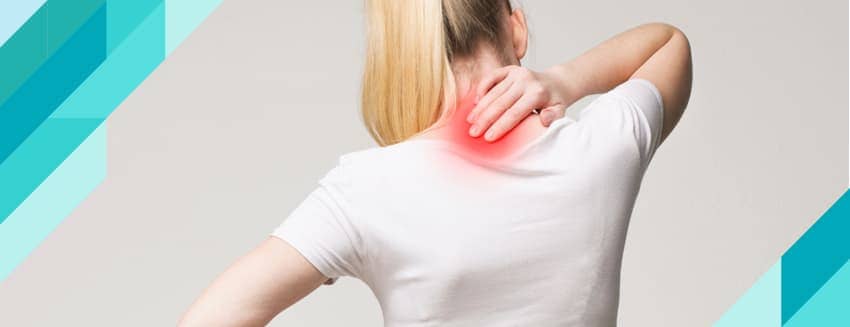
Scoliosis occurs when the spine deviates from its natural straight alignment and curves sideways; it is usually noticed during growth in childhood or adolescence. This condition can develop due to various causes, such as genetic predisposition, congenital spinal anomalies, muscle diseases, or postural abnormalities. Scoliosis can affect his posture, leading to asymmetries in his shoulders, hips, and lower back. While mild cases usually do not cause noticeable symptoms, advanced scoliosis can result in back and lower back pain, breathing difficulties, and limited mobility. Treatment varies depending on the severity and cause of the curvature; it may involve monitoring, physical therapy, specialized exercises, the use of a brace, or surgical intervention. Early diagnosis is crucial for preventing the progression of scoliosis and increasing the success of treatment.
Scoliosis encompasses a wide range of types and degrees of severity. Idiopathic scoliosis is the most common type, and its cause is generally unknown; it is typically detected during growth spurts in childhood and adolescence. Congenital scoliosis, on the other hand, develops as a result of birth defects and is usually associated with congenital deformities in the structure of the spinal bones. Additionally, neuromuscular scoliosis, which arises from muscle and nerve disorders, occurs because the muscles are unable to properly support the spine. Scoliosis is usually diagnosed through a physical examination by a doctor, along with imaging methods such as X-rays, magnetic resonance imaging (MRI), or computed tomography (CT).
What Are the Symptoms of Scoliosis (Spinal Curvature)?
The symptoms of scoliosis can vary depending on the degree and type of curvature in the spine. While scoliosis is often difficult to detect in its early stages, it becomes more apparent as it progresses, manifesting with more noticeable physical symptoms. Here are the common symptoms of scoliosis:
1. Postural Abnormalities
- Asymmetrical shoulder position (one shoulder may be higher than the other).
- Misaligned hips or one side appearing more prominent.
- The torso appears to be leaning to one side.
- A lateral curvature of the spine in an “S” or “C” shape.
2. Other Visible Signs
- One side of the waistline is more pronounced.
- Clothing not sitting properly (for example, one pant leg always appearing longer).
- One side of the rib cage appearing more prominent or further forward than the other (especially noticeable when bending forward).
3. Physical Discomforts
- Mild back or lower back pain (usually seen in advanced scoliosis).
- Limited mobility due to the curvature of the spine.
- In advanced cases, difficulty breathing when the curvature affects lung or heart function.
4. Psychological and Social Symptoms
- Lack of self-confidence, particularly during adolescence, due to changes in physical appearance.
- Shyness or timidity in social settings upon noticing a postural abnormality.
When Should You See a Doctor?
- If you notice asymmetry in your child’s shoulders, hips, or back.
- If complaints of back pain are recurring.
- If a posture problem has begun to become noticeable.
- Early diagnosis is vital for preventing the progression of scoliosis and increasing the success of treatment. Therefore, it is recommended to consult an orthopedist or physical therapist as soon as symptoms are noticed.
What Are the Types of Scoliosis (Spinal Curvature)?
Scoliosis is a condition caused by a lateral curvature of the spine and is classified into different types based on various causes, age groups, and developmental stages. Accurately identifying these types is critical in determining the course of treatment. Here are the main types of scoliosis:
1. Idiopathic Scoliosis
Idiopathic scoliosis is the most common type of scoliosis, and its cause is not fully understood. It is usually noticed during adolescence and can occur in either gender, though it is more common in females. This type of scoliosis tends to progress during the growth period. Idiopathic scoliosis is divided into three subgroups based on age:
Infantile Idiopathic Scoliosis: It develops between the ages of 0 and 3 and is rare.
Juvenile Idiopathic Scoliosis: Develops between the ages of 3 and 10 and may be detected earlier.
Adolescent Idiopathic Scoliosis: Occurs between the ages of 10 and 18 and is the most common type. Most cases are diagnosed during this period. Treatment generally varies depending on the degree of curvature; while physical therapy and monitoring may be sufficient for mild cases, the use of a brace or surgical intervention may be necessary for severe cases.
2. Congenital Scoliosis
Congenital scoliosis is a type caused by congenital factors. It results from the incomplete formation or improper fusion of the spinal bones while the fetus is in the womb. While it may be noticeable at birth, some cases are detected during the growth process. In this type of scoliosis, the curvature can progress rapidly and usually requires surgical intervention. Early diagnosis and intervention are crucial for minimizing the effects of congenital scoliosis.
3. Neuromuscular Scoliosis
Neuromuscular scoliosis develops as a result of disorders of the muscle or nervous system. Conditions such as cerebral palsy, muscular dystrophy, spinal muscular atrophy, and spinal cord injuries can cause neuromuscular scoliosis. Because of muscle weakness, the spine cannot be properly supported, which leads to curvature. In this type of scoliosis, which typically progresses rapidly, treatment options include physical therapy, the use of a brace, and surgery.
4. Degenerative Scoliosis
Degenerative scoliosis usually develops as a result of age-related wear and tear of the spinal discs, ligaments, or joints. This type is more common in adults and the elderly. Factors such as osteoporosis, arthritis, and trauma can trigger degenerative scoliosis. Symptoms include back and lower back pain, limited mobility, and postural abnormalities. Treatment generally involves pain management, physical therapy, and, in severe cases, surgical intervention.
5. Functional Scoliosis
Functional scoliosis is a temporary type of curvature that develops without any structural problem in the spine. The underlying cause is usually muscle spasms, a difference in leg length, or poor posture. This type of scoliosis can usually be completely corrected by treating the underlying cause. Treatment focuses on physical therapy and posture correction methods.
6. Syndromic Scoliosis
Syndromic scoliosis arises in association with genetic and systemic disorders such as neurofibromatosis, Marfan syndrome, or Down syndrome. This type of scoliosis is frequently seen alongside other musculoskeletal or nervous system issues. The treatment process is planned using a multidisciplinary approach and is managed in conjunction with the underlying syndrome.
Scoliosis is a complex spinal condition with various types and degrees of severity. Idiopathic, congenital, neuromuscular, degenerative, functional, and syndromic types are distinguished from one another by their unique causes and treatment approaches. Early diagnosis and regular follow-up are critical in preventing the progression of scoliosis and minimizing its effects. Therefore, when symptoms of scoliosis are noticed, an evaluation by a specialist is necessary.
How Is Scoliosis (Spinal Curvature) Recognized?
Scoliosis typically manifests as a curvature of the spine or a postural abnormality. Early diagnosis is crucial for preventing the progression of scoliosis. Both visually observable signs and medical methods are used to diagnose scoliosis. Here’s how scoliosis can be identified:
1. Visible Signs
Early signs of scoliosis include physical asymmetries and postural abnormalities. These include:
Shoulder Asymmetry: One shoulder being higher than the other.
Hip Asymmetry: The hips are misaligned, or one side appears more prominent than the other.
Tilted Torso: The person’s torso is tilted to one side, or his center of gravity appears shifted.
Rib Protrusion: One side of the ribs appearing more prominent than the other when bending forward.
Clothing Mismatch: Signs that clothing does not fit the body properly, such as one pant leg consistently appearing longer than the other.
2. Physical Tests
Here are some simple tests used by a specialist during a physical examination to detect scoliosis:
Adams Forward Bend Test: When the patient bends forward, the curvature of the spine and rib protrusion can be clearly observed. This test is used to determine the presence of scoliosis and which side it affects.
Spinal Examination: Information about the severity of the curvature is obtained by manually examining the spine.
3. Medical Imaging Methods
Medical imaging methods are used to definitively diagnose scoliosis and determine its severity:
X-ray: Used to assess the structure of the spine and measure the degree of curvature. The severity of scoliosis is evaluated using a measurement called the Cobb angle.
Magnetic Resonance Imaging (MRI): A more detailed examination is performed to check for any other issues in the spine and nerve tissues.
Computed Tomography (CT): Used to examine the bone structure of the spine in detail.
4. Complaints and Symptoms
In some cases, scoliosis can cause pain or other symptoms. These include:
- Back, lower back, or shoulder pain.
- A feeling of fatigue after standing for long periods.
- In advanced cases, respiratory problems such as shortness of breath may occur.
When Should You See a Doctor?
If your child or you experience one or more of the above symptoms—especially if you notice physical asymmetry—you should consult an orthopedic specialist. Early diagnosis is critical for halting the progression of scoliosis and ensuring effective treatment. Depending on the situation, treatment options such as physical therapy, bracing, or surgery may be recommended.
How Is Scoliosis (Spinal Curvature) Diagnosed?
1. Assessment of Medical History and Symptoms
The doctor will ask when the symptoms of scoliosis were first noticed, whether the curvature has progressed, and if there are any other complaints, such as pain or fatigue. Additionally, genetic factors are assessed by asking whether there is a family history of scoliosis.
2. Physical Examination
The first signs of scoliosis can be observed during a physical examination. Some of the tests the doctor performs include:
Adams Forward Bend Test: The patient is asked to bend forward. During this test, the spine, ribs, and back are examined. A protrusion on one side of the ribs or back may be a sign of scoliosis.
Posture Analysis: The patient’s posture, shoulders, hips, lumbar curves, and body symmetry are carefully evaluated.
Spinal Examination: The spine is palpated to determine the direction and nature of the curvature.
3. Imaging Methods
Medical imaging methods are used to definitively diagnose scoliosis and determine the degree of the curvature:
X-ray: Provides a complete view of the spine. The degree (severity) of the curvature is determined by measuring the Cobb angle. This method is the gold standard for diagnosing scoliosis.
Magnetic Resonance Imaging (MRI): Used for detailed examination of the spinal cord, nerves, and soft tissues. It is preferred especially when neuro-muscular or congenital scoliosis is suspected.
Computed Tomography (CT): Used to examine the bony structure of the spine in three dimensions. It may be necessary in more complex cases.
4. Determining the Degree of Scoliosis
The risk of scoliosis progression and the treatment plan are determined based on the degree of curvature:
Mild Scoliosis: The curvature is between 10 and 20 degrees.
Moderate Scoliosis: The curvature ranges from 20 to 40 degrees.
Severe Scoliosis: The curvature is greater than 40 degrees and typically requires surgical treatment.
5. Other Tests
If the scoliosis is caused by an underlying neurological or systemic disease, the doctor may order additional tests:
Neurological Examination: Muscle strength, reflexes, and nerve function are assessed.
Blood Tests: These can be used to identify conditions associated with other systemic diseases in cases of congenital or syndromic scoliosis.
The Importance of Early Diagnosis
Early diagnosis of scoliosis is crucial for controlling the risk of progression and implementing appropriate treatment methods. Regular checkups, especially for children who are still growing, ensure that scoliosis is detected in its early stages and increase the likelihood of successful treatment. Therefore, it is recommended to consult an orthopedic specialist if any postural abnormalities or physical asymmetries are noticed.
Scoliosis (Spinal Curvature) Grades and Classification
Scoliosis is graded and classified based on the severity and structure of the spinal curvature. This grading is critical for determining the risk of scoliosis progression and selecting appropriate treatment methods. The curvature is typically measured by calculating the Cobb angle on X-ray images. Here are the grades and classifications of scoliosis:
1. Scoliosis Grades
Scoliosis is classified as follows based on the Cobb angle:
Mild Scoliosis (10–20 degrees): The curvature of the spine is usually difficult to detect and generally does not cause pain. Regular monitoring and physical therapy are recommended as treatment. In most cases, surgical intervention is not required.
Moderate Scoliosis (20–40 degrees): The curvature becomes more pronounced, and physical symptoms such as shoulder and hip asymmetry are visible. Treatment may include the use of a brace and physical therapy. If the curvature progresses, surgery may be considered.
Severe Scoliosis (40 degrees and above): The curvature has reached a serious level and can often lead to pain, limited mobility, and even breathing problems. Scoliosis of this severity generally requires surgical intervention.
Very Severe Scoliosis (80 degrees and above): In this rare condition, spinal curvature can cause serious health problems. It can particularly affect heart and lung function. It almost always requires surgical treatment.
2. Classification by Curvature Shape
C-Shaped Scoliosis: This is a condition in which the spine curves to one side. It is generally seen in mild cases.
S-Shaped Scoliosis: A more complex type in which the spine curves in two different areas. It usually occurs in progressive cases and is more difficult to treat.
3. Classification by Age of Onset
Infantile Scoliosis (ages 0–3): It appears in the first few years after birth. The curvature may sometimes correct itself, but there is a risk of rapid progression.
Juvenile Scoliosis (ages 3–10): Occurs during childhood and can progress rapidly during growth spurts. Early intervention is important.
Adolescent Scoliosis (ages 10–18): Develops during growth spurts in adolescence. It is the most common type of scoliosis and requires regular follow-up.
Adult Scoliosis (age 18 and older): May result from the progression of scoliosis that began in childhood or adolescence, or from degenerative changes associated with aging.
4. Classification by Cause
Idiopathic Scoliosis: A type of scoliosis with an unknown cause; it is the most common form.
Konjenital Skolyoz: It is caused by spinal anomalies present at birth.
Neuromuscular Scoliosis: It results from muscle or nerve disorders.
Degenerative Scoliosis: This type results from changes in the structure of the spine associated with aging in adults.
5. Classification Based on Progression
Progressive Scoliosis: The curvature worsens over time. This type is more common during childhood and adolescence.
Non-Progressive Scoliosis: The curvature remains stable and generally does not require additional treatment.
The severity and classification of scoliosis are crucial for developing an appropriate treatment plan. Early diagnosis and regular follow-up play a critical role in controlling the progression of the curvature. While physical therapy and exercises may be sufficient for mild cases, severe cases may require surgical intervention. Therefore, if scoliosis is suspected, a specialist should be consulted.
Scoliosis Treatment: How Is Spinal Curvature Corrected?
Scoliosis treatment is planned based on the degree of the curvature, the risk of progression, age, and overall health. The goal of treatment is to correct the curvature, halt its progression, and improve the person’s quality of life. Here are the methods for treating scoliosis:
1. Observation and Regular Follow-Up
Mild Scoliosis (10–20 degrees): If the curvature is mild and there is no risk of progression, the doctor will recommend regular follow-up. Since scoliosis can progress during the growth period in children and adolescents, X-rays are typically taken every 4–6 months to monitor the spine.
Exercises: For mild scoliosis, exercises that strengthen the muscles and support posture may be recommended.
2. Physical Therapy and Rehabilitation
The Schroth Method: The Schroth Method, a physical therapy technique developed specifically for scoliosis, aims to correct spinal curvature and restore muscle balance. This method includes breathing techniques and postural exercises.
Pilates and Yoga: These can help correct postural issues and increase muscle flexibility. However, these exercises should be performed under professional supervision.
3. Brace Therapy
In cases where the curvature is between 20 and 40 degrees, the use of a brace is recommended to prevent the progression of scoliosis, particularly in children who are still growing.
Purpose of the Brace: Braces stop the progression of the curvature by keeping the spine in the correct position, but they do not correct the existing curvature.
Types of Braces:
Boston Brace: It tightly wraps around the torso and supports the spine.
Chêneau Brace: A specialized brace that provides three-dimensional scoliosis correction.
Milwaukee Brace: Used in more severe cases and features shoulder support.
4. Surgical Treatment
When the curvature exceeds 40–50 degrees and continues to progress, surgical intervention is generally necessary. Surgery is also recommended when severe scoliosis affects respiratory or cardiac function.
Types of Surgery:
Spinal Fusion: A procedure that uses metal rods, screws, and hooks to fuse the vertebrae together and stabilize the spine in a fixed position.
Growth-Adjustable Rods: Special rods used in children that can be adjusted at specific intervals to avoid hindering growth.
Vertebral Body Tethering (VBT): A minimally invasive method that aims to correct the spine using a flexible strap while preserving mobility.
5. Lifestyle and Supportive Approaches
Posture Correction: Proper sitting, standing, and lifting habits in daily life can help protect the spine.
Pain Management: Pain can be managed through physical therapy, massage, hot/cold therapy, and, in some cases, pain medication.
6. Alternative Treatments
Some individuals may try acupuncture, manual therapy, or chiropractic care as complementary approaches. However, these methods have not been scientifically proven to halt the progression of scoliosis and should not be used without professional advice.
Scoliosis treatment is tailored to the degree of the curvature and the individual’s specific circumstances. While observation and physical therapy may be sufficient in mild cases, surgical intervention may be necessary in advanced cases. Early diagnosis and proper treatment planning are critical for managing the effects of scoliosis and improving the individual’s quality of life. If scoliosis is suspected, an orthopedic specialist should be consulted.
Scoliosis (Spinal Curvature) Surgery
Scoliosis surgery is a surgical procedure performed to correct the curvature of the spine, halt its progression, and stabilize the spine. It is generally recommended when the curvature exceeds 40–50 degrees or when the curvature negatively affects quality of life. Here’s what you need to know about scoliosis surgery:
1. Who Needs Surgery?
Severe Curvature: Cases where the Cobb angle exceeds 40–50 degrees.
Rapidly Progressing Scoliosis: Rapid progression of the curvature, particularly in growing children.
Physical Problems: Severe scoliosis that affects respiratory or cardiac function.
Pain and Limited Mobility: Pain, difficulty with physical activities, and postural abnormalities.
Cosmetic Concerns: A noticeable curvature of the spine that significantly affects appearance.
2. Types of Surgery
Spinal Fusion: This is the most common surgical method for treating scoliosis.
The curved vertebrae are stabilized using metal rods, screws, and hooks, and these vertebrae are fused together. The goal is to realign the spine and make it stable.
Growth-Adjustable Rods:
This method is used in children while they are still growing. Flexible rods are lengthened at specific intervals to correct the curvature.
Vertebral Body Tethering (VBT):
This is a minimally invasive technique. A flexible band is placed along the spine to correct the curvature as the vertebrae grow naturally.
Spinal mobility is preserved, making this method particularly preferred for young patients.
3. Surgical Process
Preparation: A detailed examination and imaging are performed before surgery. The patient’s overall health is assessed.
Surgery Duration: The procedure typically lasts between 4 and 8 hours.
Anesthesia: The procedure is performed under general anesthesia.
Surgical Technique: The surgeon stabilizes the spine by placing rods and screws in the areas to be corrected. Bone grafts are used for fusion.
4. Postoperative Process
Hospital Stay: The hospital stay usually lasts 5–7 days.
Recovery Process: The postoperative recovery process varies between 6 and 12 months. Caution is required during daily activities for the first few weeks.
Physical Therapy: Physical therapy is recommended to improve mobility and strengthen the spine.
Pain Management: Postoperative pain can be managed with medications prescribed by your doctor.
5. Risks of Surgery
As with any surgical procedure, scoliosis surgery carries certain risks:
Infection: The risk of infection at the surgical site.
Bleeding: Blood loss may occur during surgery.
Nerve Damage: Although rare, nerve damage is a serious risk.
Fusion Problems: Additional surgery may be required if the vertebrae do not fuse properly.
6. Benefits of Surgery
- Proper alignment of the spine.
- Halting the progression of the curvature.
- Reduction or complete elimination of pain.
- Improved aesthetic appearance and posture.
- Restoration of normal respiratory and cardiac function.
7. Life After Surgery
Return to Normal Activities: Most patients can return to their normal activities within 6 months. However, strenuous physical activities should be avoided.
Long-Term Follow-Up: Regular follow-up visits after surgery are important. The positioning of metal implants and the condition of the spine are monitored regularly.
Conclusion
Scoliosis surgery is an effective and permanent method for treating severe spinal curvatures. Appropriate patient selection, surgery performed by an experienced surgeon, and regular follow-up increase the success of treatment. The decision to undergo surgery as part of the scoliosis treatment process should be evaluated based on the patient’s individual needs.
Things Patients Should Keep in Mind After Scoliosis (Spinal Curvature) Surgery
The recovery process following scoliosis surgery is extremely important for ensuring the success of the surgery and improving the patient’s quality of life. During this process, it is essential to follow the doctor’s recommendations and be mindful of certain habits to prevent complications and ensure the spine heals properly. Here are the key points patients should keep in mind:
1. Physical Activity
Light Exercises: Light activities such as walking are recommended during the first few weeks. This improves blood circulation and supports healing.
Avoiding Heavy Lifting: For the first 3–6 months, he should avoid heavy lifting or movements that strain the spine.
Posture: Develop good posture habits and avoid bending or twisting movements that place excessive strain on the spine.
Physical Therapy: A physical therapy program may be started as recommended by his doctor. This helps strengthen muscles and improve mobility.
2. Daily Life and Activities
Lying Down and Getting Up: When getting out of bed after surgery, he should use the “roll-over” technique to avoid straining his spine.
Sitting and Standing: It is important to sit on firm surfaces and to be careful when sitting down and standing up. He should move slowly, using the chair for support.
Driving: He should avoid driving for the first few months. During travel, he should avoid sitting for long periods and take breaks to move around as needed.
3. Nutrition
Focus on Bone Health: During the recovery process, a diet rich in calcium, vitamin D, and protein should be prioritized.
Weight Management: Since excess weight places additional strain on the spine, weight should be managed through a balanced diet.
Water Intake: It is important to drink an adequate amount of water to support the body’s overall recovery.
4. Care of the Surgical Site
Hygiene: Dressings should be changed regularly and the wound kept clean to prevent infection of the surgical site.
Checking the Stitches: If signs of infection—such as redness, swelling, or discharge—appear around the stitches, consult a doctor immediately.
Bathing: Generally, care should be taken not to get the area wet for the first few weeks after surgery. He may take a shower once his doctor gives permission.
5. Pain Management
Pain Relievers: Take the pain relievers prescribed by his doctor as directed. However, he should be careful not to exceed the recommended dosage.
In Case of Discomfort: If he experiences prolonged or severe pain, he should consult his doctor.
6. Sleep Habits
Sleeping Position: Sleeping on his back or side is best during the first few months. A firm, orthopedic mattress is recommended.
Pillow Use: A suitable pillow that supports the head, neck, and spine should be used.
7. Returning to Social and Professional Life
Returning to School or Work: Depending on the postoperative recovery process, he can generally return to jobs requiring light physical activity within 6–8 weeks. For more strenuous work, he should wait for his doctor’s approval.
Sports and Hobbies: Contact sports or strenuous physical activities should be avoided for 6 months following surgery. Light exercise may be started upon the doctor’s recommendation.
8. Regular Follow-Up Visits
Doctor Follow-up: He should attend regular doctor’s appointments throughout the first year after surgery. X-rays will be taken to assess the condition of the spine.
Long-Term Monitoring: The condition of the metal implants and the fusion should be monitored regularly.
A successful recovery process after scoliosis surgery depends on the patient moving carefully and following the doctor’s recommendations. Engaging in physical activities in moderation, maintaining hygiene in the surgical area, and adopting healthy lifestyle habits support proper healing of the spine. In the event of any complications, the doctor should be consulted without delay.
Innovations in Scoliosis Surgery
New techniques and technologies developed in scoliosis surgery offer less invasive approaches, making the treatment process easier for patients and shortening recovery times. Here are some important innovations in scoliosis surgery:
1. Vertebral Body Tethering (VBT) - Tethered Scoliosis Surgery
VBT is a minimally invasive method that corrects the curvature by utilizing the spine’s natural growth process. The curvature is gradually corrected using a flexible tether placed on the front of the spine. This method is particularly preferred for young patients who still have growth potential and preserves spinal mobility to a large extent. Compared to traditional surgical methods, it preserves mobility without the need for spinal fusion.
2. Magnetically Controlled Growing Rods (MCGR)
Magnetically controlled growing rods, a method designed for pediatric scoliosis patients, are implanted into the spine during surgery. The rods are extended using an external magnetic device to adapt to the spine’s growth process. This method eliminates the need for repeated surgeries, thereby improving patient comfort and allowing for controlled correction of the curvature during the growth process.
3. Minimally Invasive Surgical Approaches
Thoracoscopic Spinal Surgery (VATS): This technique, which allows access to the spine through small incisions, results in less pain and a shorter recovery period. Particularly preferred for scoliosis surgeries performed in the chest area, this method also ensures greater aesthetic satisfaction for patients.
4. Robotic-Assisted Surgery
Robotic surgical systems enhance precision in spinal surgery. With robotic assistance, surgeons can place spinal screws more accurately, which increases the success rate of the surgery while reducing the risk of complications. It is noted that robotic surgery offers a more reliable solution, particularly in complex scoliosis cases.
5. 3D Printing Technology
In scoliosis surgery, implants produced using patient-specific 3D printing technology are used. This technology enables surgeons to provide solutions tailored to each patient’s spinal anatomy. Models produced via 3D printing also facilitate preoperative planning.
These innovations in scoliosis surgery are making treatment processes more effective and patient-friendly. Less invasive approaches preserve spinal mobility while shortening the recovery process. The choice of method depends on factors such as the patient’s age, the severity of the scoliosis, and overall health. Achieving successful outcomes in scoliosis treatment with innovative surgical methods is now more feasible than ever.
How Should Scoliosis Exercises Be Performed?
Scoliosis exercises are physical therapy methods used to control spinal curvature, strengthen muscles, and correct posture. When performed regularly and correctly, these exercises can slow the progression of scoliosis and support spinal health. Here are some important facts and examples regarding scoliosis exercises:
1. The Purpose of the Exercises
Achieve Muscle Balance: Strengthen weak muscles and relax tight muscles.
Correcting Posture: Aligning the spine as much as possible.
Increasing Spinal Flexibility: To improve range of motion and prevent postural abnormalities.
Reducing Pain: Alleviating muscle tension and pressure on the spine.
2. Precautions to Take Before Starting the Exercises
Professional Guidance: Consult a doctor, physical therapist, or scoliosis specialist before beginning any exercises.
Exercise Selection: Exercises appropriate for the severity and type of scoliosis should be identified.
Consistency: It is important to perform the exercises regularly for them to be effective.
3. Recommended Exercises for Scoliosis
a. Breathing Exercises (Schroth Method): This method, used in the treatment of scoliosis, focuses on strengthening asymmetrical muscle groups and aligning the spine.
The goal is to expand the rib cage and lengthen the spine by taking deep breaths.
How to Do It: Sit upright, take a deep breath toward the side of the curve, and exhale slowly.
b. Cat-Cow Stretch: This is ideal for stretching and relaxing the spine.
How to do it: Get on your hands and knees. As you inhale, arch your back (cat pose); as you exhale, lower your back and lift your head (cow pose).
c. Side Stretch: Increases spinal flexibility and strengthens the muscles.
How to Do It: Stand upright. Place one hand on top of your head and stretch sideways. Repeat on both sides.
d. Bridge Pose: Helps strengthen the lower back muscles.
How to do it: Lie on your back, bend your knees, and place your feet flat on the floor. Slowly lift your hips, hold for a few seconds, then lower them.
e. Plank: Strengthens the abdominal and back muscles and supports the spine.
How to Do It: Lie face down on the floor and rest on your elbows and toes. Keep your body in a straight line and hold this position for 20–30 seconds.
4. Things to Avoid During Exercise
Strenuous Exercises: Avoid exercises that are excessively strenuous or high-impact.
Asymmetrical Movements: Avoid one-sided movements that could worsen scoliosis.
Sudden Movements: Avoid movements that place sudden stress on the spine.
5. Duration and Frequency of Exercises
Exercises should generally be performed 3–5 days a week. Each exercise set can consist of 10–15 repetitions. Duration and intensity should be adjusted according to the physical therapist’s recommendations.
Scoliosis exercises are an effective method for preventing the progression of scoliosis and strengthening the muscles. However, it is important that these exercises be performed using proper technique and under the guidance of a specialist. Through regular exercise, spinal health can be maintained, postural problems can be prevented, and pain can be reduced.
How Should People with Scoliosis Sleep?
It is important for people with scoliosis to adopt the correct sleeping positions to alleviate pain by reducing pressure on the spine and to improve sleep quality. Incorrect sleeping positions can further worsen the curvature of the spine or cause discomfort. Here are the correct sleeping positions for people with scoliosis and what to keep in mind:
1. Side-Lying Position (The Most Ideal Position)
The most suitable sleeping position for people with scoliosis is generally lying on their side.
Depending on the shape of the curvature, one side may be preferred to support the spine:
C-shaped scoliosis: Sleeping on the inner side of the curve can help keep the spine in a more comfortable position.
For S-shaped scoliosis: appropriate pillows and supports should be used to balance both curves of the spine.
Use of Supports: Placing a pillow between the knees helps align the hips and spine, thereby reducing pressure.
2. Sleeping on Your Back
Sleeping on his back is also a suitable option for people with scoliosis, as this position helps keep the spine in its natural alignment.
Pillow Under the Knees: Placing a pillow under the knees reduces pressure on the lower back and promotes a more restful sleep.
Using a Firm Mattress: Choosing an orthopedic mattress supports the spine when sleeping on his back.
3. Positions to Avoid
Sleeping on Your Stomach: Sleeping on your stomach applies asymmetrical pressure to the spine and may cause scoliosis to worsen. It can also create tension in the neck and back.
Crooked or Twisted Positions: Sleeping positions that cause the spine to bend in an incorrect position should be avoided.
4. Mattress and Pillow Selection
Orthopedic Mattress: A medium-firm mattress that supports the spine should be chosen. Very soft mattresses can increase spinal curvature, while very firm mattresses can cause discomfort.
Pillow: A pillow that maintains the natural alignment of the head and neck should be used. It is important that the pillow is neither too high nor too low.
Body Pillow: A long pillow can be used to support the entire body when sleeping on his side.
5. The Importance of the Sleep Environment
A Flat Surface: The bed should be flat and provide adequate support for the body.
A Comfortable Sleep Environment: A sufficiently dark, quiet, and comfortable sleep environment makes it easier for people with scoliosis to rest.
Proper Sleep Duration: Adequate, high-quality sleep supports scoliosis treatment and helps alleviate pain.
The sleeping position of scoliosis patients should be such that it minimizes pressure on the spine, thereby reducing pain and protecting spinal health. Sleeping on one’s side or on one’s back are generally the most suitable options. Choosing an orthopedic mattress and an appropriate pillow helps ensure proper spinal alignment while improving sleep quality. It may be helpful to consult a physical therapist or doctor for detailed recommendations regarding sleeping positions.
Is Scoliosis Surgery Recommended for Children?
Scoliosis surgery in children is generally preferred when the curvature has reached a severe degree or is progressing rapidly. However, the decision to undergo surgery depends on the child’s age, the degree of spinal curvature, and the impact of scoliosis on the child’s overall health. Surgery is performed to correct the spinal curvature, halt its progression, and improve the child’s quality of life. Here are the details regarding scoliosis surgery in children:
1. When Surgery Is Necessary
Severe Curvature: Surgery is considered when the Cobb angle exceeds 40–50 degrees.
Rapid Progression: Rapid progression of scoliosis during the child’s growth period.
Physical Discomfort: When the spinal curvature causes pain, limited mobility, or postural abnormalities.
Impact on Organ Function: When the curvature affects heart and lung function, leading to respiratory problems.
2. Surgical Methods Used in Children
Growing Rods: Preferred for younger children. Rods that allow the spine to grow are implanted and lengthened at specific intervals as the child grows.
Thanks to magnetically controlled rods (MCGR), the rods can be lengthened using an external device without the need for repeated surgery.
Spinal Fusion: A common method used to correct scoliosis and stabilize the spine. It is preferred for older children or in cases where growth is largely complete.
Vertebral Body Tethering (VBT): This is a minimally invasive technique that preserves spinal mobility. The curvature is corrected and monitored as the child grows using a flexible tether placed in the spine.
3. Benefits of Surgery
Positioning the spine in the optimal alignment for correction. Halting the progression of the curvature. Correcting posture and reducing pain. Preserving organ function and resolving respiratory problems. Allowing the child to participate more comfortably in social and physical activities.
4. Risks and Challenges
As with any surgical procedure, scoliosis surgery carries certain risks:
Infection: The risk of infection at the surgical site.
Bleeding: Bleeding may occur during or after surgery.
Fusion Problems: The vertebrae may not fuse as expected.
Nerve Damage: Although rare, damage to the nerves surrounding the spine is possible. These risks can be minimized when the surgery is performed by experienced surgeons.
5. Factors to Consider When Deciding on Surgery
Child’s Age: Surgical intervention is planned with the child’s growth potential in mind.
Degree of Scoliosis: In mild and moderate cases, non-surgical methods (braces, physical therapy) are generally preferred.
Informing the Family and Child: The surgical process, risks, and expectations should be discussed in detail with the family and the child.
Scoliosis surgery is an effective option for children with severe curvature when other treatment methods are insufficient. Early diagnosis and proper planning increase the likelihood of a successful surgery and improve the child’s quality of life. It is important to consult with an orthopedic specialist or a surgeon experienced in scoliosis when making the decision to proceed with surgery.