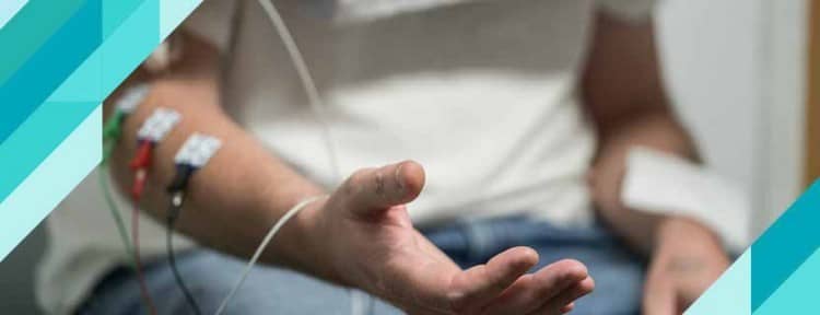
Electromyography (EMG) is the process of measuring the responses of muscles to electrical signals sent by nerves. This test is used to help detect abnormalities in the neuromuscular (muscle and nerve) system. During an EMG, one or more thin needle electrodes are inserted into muscle tissue. The electrical activity collected by the electrodes is then displayed on an oscilloscope screen. Normally, muscles do not produce electrical signals while at rest, but activity may be observed for a short time after the electrodes are inserted. Electrical activity is measured and evaluated during rest, light contraction, and strong contraction of the muscles.
In addition, during an EMG test, the patient may be asked to move his muscles in specific ways. This is done to observe and evaluate how the muscles respond. In addition to EMG, a nerve conduction study (NCS) may also be performed. NCS measures the speed and magnitude of electrical signals transmitted by nerves. By using EMG and NCS together, the presence, location, and extent of nerve and muscle damage can be determined. These procedures play a crucial role in the diagnosis and treatment of many nerve and muscle disorders.
Advantages of Having an EMG (Electromyography) Test Performed by a Neurologist
The quality of the EMG device and its software for electrical noise separation and filtering are extremely important for precise signal monitoring and testing. Our hospital uses state-of-the-art
EMG devices that are among the world’s leading technologies in this field. However, these tests are typically performed by technicians and interpreted by specialist physicians. This situation can lead to some diagnoses being missed. For this reason, at our hospital—a practice rarely seen in our country—EMG tests are performed exclusively by a “Neurologist.”
For Which Conditions Is an EMG (Electromyography) Test Performed?
The EMG test is used to diagnose disorders affecting the structure and function of muscles and nerves. The functionality of muscles and the nervous system is determined through the EMG test. Among these conditions are neuromuscular disorders, ALS, spinal cord disorders, and focal neuropathies.
The EMG test is most commonly
used to diagnose the following conditions:
Focal Neuropathies
EMG (electromyography) is generally used to diagnose problems that cause damage to a single nerve. Common examples include carpal tunnel syndrome in the wrist and cubital tunnel syndrome in the elbow.
In
addition, EMG is used in cases of peroneal nerve neuropathy in the leg or tarsal tunnel syndrome in the ankle. In clinical practice, sciatic nerve damage may occur due to improper injections in the hip or hip dislocations. In such cases, an EMG test is the preferred method for establishing an accurate diagnosis. Furthermore, this method provides information regarding the severity of nerve damage and the prognosis for recovery in cases of facial paralysis.
Diabetes and Kidney Failure
EMG is used for diagnostic purposes
in cases of widespread peripheral nerve damage caused by diabetes, kidney failure, and certain genetic factors that impair nerve function.
Damage
to Nerve Roots Due to Lumbar and Cervical Disc Herniations It is also used to identify diseases that cause damage to the nerve roots responsible for lumbar or cervical disc herniations originating from the spinal cord.
Myopathies
The EMG test is an effective
method for diagnosing diseases that cause damage and degeneration of muscle fibers.
Polio or ALS The EMG method is also used to diagnose diseases that cause damage to motor nerve cells in the spinal cord, such as polio or ALS
.
Neuromuscular
Diseases
EMG is an important test
used to diagnose diseases—such as myasthenia gravis—in which electrical transmission between nerves and muscles is impaired.
Although the EMG procedure may vary depending on the patient’s condition and the treating physician’s preferences, the general steps are as follows:
Pre-Procedure Preparations:
- Your doctor will explain the details of the procedure and give you the opportunity to ask questions.
- In most cases, you do not need to follow a special diet before the test.
- You should inform the doctor of all medications and supplements you are taking.
- On the day of the procedure, avoid applying lotion or oil to your skin.
- If you have a pacemaker, you must inform your doctor.
Procedure:
- The procedure is performed by a neurologist. During the test, thin, sterile needles are inserted into the muscle. These needles measure electrical activity and display it on an oscilloscope.
- The insertion of the needles is generally painless, though he may feel a slight discomfort.
- The patient is asked to perform various movements by contracting his muscles. This provides information about how well the nerves stimulate the muscles and how the muscles respond to these stimuli.
Post-Procedure Instructions:
- Mild muscle soreness may occur after the procedure. If he experiences any severe pain, swelling, or pus, he should contact his doctor.
The “NCS” Procedure
NCS (Nerve Conduction Studies) is a test that measures how quickly and effectively electrical impulses are transmitted along a nerve. This procedure is typically performed in conjunction with an EMG test, and together, both procedures help diagnose conditions such as nerve damage and muscle disorders.
Before the Procedure:
- The NCS procedure generally does not require any special preparation.
- Just as with an EMG, all medications taken should be reported to the doctor, and the skin should be kept clean on the day of the procedure.
During the Procedure:
- During the NCS, mild electrical impulses are delivered via electrodes placed on specific nerves. This measures the nerve’s response to electrical stimulation.
- He may feel slight discomfort during the test, but it is usually brief and mild.
After the Procedure:
- After the test, the gel used to attach the electrodes is removed, and patients can usually resume their normal activities.
- Both tests are important tools for assessing nerve and muscle health and are used in the diagnosis of many neuromuscular disorders.


