
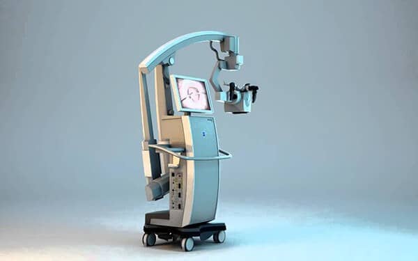
The tumor fluorescence-guided surgical microscope is a type of microscope that uses special fluorescent dyes to make tumor tissue prominent during surgery. This technology makes it easier to distinguish tumor tissue from healthy tissue, enabling surgeons to perform more precise and effective interventions.
Microscope Features
Fluorescent Staining Technology: Fluorescent dyes make the tumor tissue prominent during surgery. These dyes glow under the microscope light, allowing surgeons to clearly see the tumor margins.
High-Resolution Imaging: The microscope provides detailed and clear images using high-resolution optical systems. This makes it possible for surgeons to see even the finest details.
Real-Time Imaging: Imaging is performed in real-time during surgery. This allows surgeons to make instant decisions and perform procedures with precision.
Integrated Imaging Systems: The microscope operates integrated with PACS (Picture Archiving and Communication System) and other clinical systems, ensuring that the obtained images are instantly recorded and analyzed.
Ergonomic Design: It is ergonomically designed to allow surgeons to work comfortably for long periods. The adjustable features of the microscope ensure that the surgeon works in the optimal position.
Microscope Benefits
Surgical Precision and Efficacy: Fluorescent staining technology ensures the clear delineation of tumor margins. This helps surgeons completely remove the tumor without damaging healthy tissues.
Reduced Risk of Complications: The clear identification of tumor tissue reduces the risk of postoperative complications. The preservation of healthy tissues accelerates the patients' recovery process.
Shorter Surgery Duration: Precise imaging and clear identification enable surgeons to perform procedures faster and more safely. This shortens the duration of the surgery and reduces the time patients remain under anesthesia.
Advanced Education and Research: The recorded images can be used as educational material for medical students and surgeons. Additionally, they constitute an important data source in clinical research.
Areas of Use
Brain Tumors: Provides precise and clear imaging during the surgical removal of brain tumors.
Orthopedic Oncology: Used in the surgical intervention of bone tumors.
General Surgery: Used effectively in the removal of liver, pancreas, and other organ tumors.
